AI for Digital Pathology
Key Aspects for Successful Delivery
Custom software engineering and quality and information-security standards, INNERLUXES helps clinical and research-focused digital pathology product companies engineer AI features for existing platforms and build AI-powered digital pathology software from scratch.
Contributor

Senior Solution Architect, Healthcare, INNERLUXES
AI for Digital Pathology: Essence
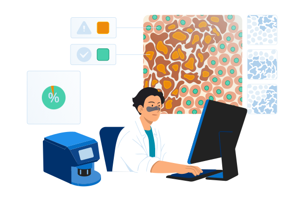
Digital pathology software with AI uses machine learning and computer vision to process whole-slide images (WSI) and identify, measure, and classify tissue patterns. In clinical settings, it helps pathologists review cases faster, quantify biomarkers more consistently, and prioritize high-risk slides. In research, it helps turn slide data into structured insights for cohort analysis, biomarker exploration, and multimodal studies.
AI in Pathology Market
The global AI in pathology market size is projected to reach USD 1.15 billion by 2033, growing at a CAGR of 27.18%. The key drivers of AI’s growth in digital pathology are the rising number of cancer and chronic disease cases, which create more slide-review work for labs, the need to process slides faster and boost productivity, the ongoing digitization of pathology workflows, and AI becoming more advanced and affordable to deploy.
How AI for Digital Pathology Works
Common use cases
AI for clinical pathology workflows

Triage and case prioritization
AI assigns a risk or priority score to each slide and pushes the most suspicious cases to the top of the worklist, so pathologists review higher-risk or likely positive cases first.

Cancer detection support
AI highlights suspicious regions on pathology slides (heatmaps or overlays), including possible metastases in lymph nodes, to guide the reviewer’s attention and reduce time spent searching.

Cancer grading and clinically important feature detection
AI detects patterns linked to severity (e.g., grading-related features) and provides structured suggestions for the pathologist’s confirmation.

Tumor segmentation and quantitative measurements
AI outlines tumor regions and calculates metrics, such as tumor area percentage, to make measurements faster and more consistent.

Biomarker and immunohistochemistry (IHC) quantification
AI counts stained cells and estimates biomarker scores (e.g., Ki-67) to speed up, standardize, and reduce the labor intensity of routine pathology scoring.

Slide and scan quality control
AI detects focus and artifact issues early (folds, bubbles, staining problems) so slides can be rescanned or routed before they affect review results or AI accuracy.

Second-read support
AI provides a secondary review layer by flagging potentially missed regions or discrepant findings, helping pathologists reduce oversight risk.
AI for research and discovery

Biomarker exploration and validation
AI helps identify image-based features associated with biomarkers, treatment response, or disease progression, supporting biomarker hypothesis generation and validation.

Cell and tissue quantification
AI classifies tissue types and counts specific cell populations across large slide sets, turning images into structured data for quantitative phenotyping, research, and trials.

Tumor microenvironment analysis
AI identifies and quantifies immune cells and spatial relationships between tumors and surrounding tissue to support biomarker research and precision oncology.

Cohort discovery and stratification
AI groups slides or patients by shared morphological patterns, biomarkers, or risk signals, helping research teams build cohorts for retrospective studies, translational research, and clinical trials.

Multimodal analysis
AI links pathology image features with genomics, outcomes, or trial data to uncover stronger disease signatures and support research-grade model development.
Reference Architecture for an AI-Powered Digital Pathology Platform
Below, INNERLUXES’ solution architects outline a reference architecture for a clinical or lab digital pathology product with AI, including how the core pathology workflow can be implemented and how the AI component can be continuously monitored, evaluated, and updated. For research-focused solutions, the architecture keeps a similar overarching logic but adds a dedicated lane for data de-identification and governance, feature-extraction storage, cohort building and search, multimodal data joins with genomics, outcomes or trial data, and an experiment workspace or research API.
Core workflow. When a scanner creates a whole-slide image (WSI) and sends it to the platform, the platform validates the file, stores it, creates or updates the case record, and routes the slide for AI analysis, if configured. Inside the AI subsystem, the slide goes through image quality checks, model selection, inference, and post-processing, which converts raw model outputs into usable results such as classifications, heatmaps, scores, and measurements. These results are stored with versioning and traceability for the model, inputs, and outputs. The case worklist and viewer are then updated so AI results and overlays are available during review, and prioritization signals can be reflected in the workflow. The pathologist opens the case, reviews the slide with or without AI support, and renders the final interpretation. After sign-out, the platform generates the report, updates the case status, and sends results to connected systems such as the LIS or LIMS.
AI lifecycle. Reviewer feedback on AI outputs is captured and stored for later analysis. The product team reviews this feedback, identifies hard cases and failure patterns, and selects relevant cases for dataset updates. After curation and annotation, the updated data is added to versioned training and evaluation datasets. The model is then retrained and tested against predefined acceptance criteria. Approved model versions are packaged and released for deployment, while production monitoring tracks performance, drift, and operational issues over time.
Security and audit layer. A cross-cutting control spans the entire platform. It verifies every user through secure sign-in, controls what each role is allowed to access or do, and encrypts data both in transit and at rest. It keeps an immutable audit trail of all important actions — slide access, AI runs, outputs, and final pathologist decisions — and supports compliance with healthcare and data-protection requirements such as HIPAA, SOC 2, and GDPR.
Keep AI as a separate back-end component instead of building it directly into the slide viewer or case-management system. This one decision solves three problems at once: you can update models without touching the pathologist workflow, run old and new model versions side by side for validation, and swap your entire inference back end (local GPU today, cloud tomorrow) without affecting users.
More importantly, before spending months on the inference pipeline itself, invest two weeks into a structured correction-capture mechanism in your viewer. Every time users correct, override, or refine AI output, that disagreement should be stored with the slide reference, the model version, and the original prediction — because those corrections are your most valuable asset for making the next model version better.
Selected Medical Imaging and Diagnostic Software Projects by INNERLUXES
Technologies We Use to Build AI Products for Digital Pathology
From pathology foundation models to the deployment stack — we assemble the toolset around your use case, data, and compliance needs.
Pathology AI and model development
 UNIVirchowCONCHPhikon
 CLAMABMIL
 U-NetHoVer-Net
 ResNetEfficientNetVision Transformer (ViT)
 PyTorch
 TensorFlow
 Keras
Keras MONAI
MONAI OpenCV
 Scikit-learn
 OpenSlide
 Azure Machine Learning
 Amazon SageMaker AI
 Google Vertex AI
 Hugging Face
 MLflow
Product engineering and deployment
 Amazon Web Services
 Microsoft Azure
 Docker
 Kubernetes
 Helm
Helm Python
 TypeScript
 C#
C# C++
C++ PostgreSQL
 Redis
Redis Apache Kafka
 Prometheus
 Grafana
Best Practices for Developing AI Products for Digital Pathology
Define a narrow, lower-risk use case before scaling the model
Instead of starting with a broad, high-risk goal such as full cancer grading or complex biomarker scoring, begin with a narrower workflow — scan QC, tissue or region-of-interest (ROI) detection, or second-read triage. These use cases are easier to validate, fit more naturally into real workflows, and help teams build evidence and user confidence. Define the intended use and regulatory posture early, because that decision shapes the data strategy, validation burden, workflow design, and product claims.
Add AI to existing software as a modular workflow layer
Many digital pathology products already have a viewer, storage layer, and core case workflow, but lack the controls needed to introduce AI cleanly — triage queues, labeling workflows, role-specific review views, and admin controls for thresholds and exceptions. Add AI as a modular layer rather than rebuild the product: separate inference services, model management, overlays or heatmaps, and review workflows. This lets you release AI features faster and iterate without destabilizing the core platform.
Make “human-in-the-loop” explicit with safe UX and auditability
AI adoption stalls when pathologists do not trust outputs or cannot explain them. Design your software so the human decision is the center: show heatmaps and regions together with confidence scores, provide “why flagged” context, and support easy overrides (confirm, reject, defer). Every AI output should be traceable — model version, data version, thresholds used, and the final human action. This is also the foundation for audits, clinical validation packages, and future regulated pathways.
Design for variability and drift from day one
“It worked in the lab” often fails in production because stain protocols, tissue prep, scanners, compression, and focus artifacts vary across sites. Treat variability as a first-class requirement: validate on multi-site data, track performance by scanner, stain, or subtype, and deploy drift monitoring. When confidence drops or drift is detected, route cases to a human-first path and trigger an active-learning loop to capture hard cases, label them, and retrain with controlled releases instead of silent model degradation.
Build evaluation like a product feature
Structure AI evaluation so every model release is tested the same way and results can be compared over time. Build a versioned, automated evaluation pipeline with fixed dataset splits, ground-truth provenance, cohort-level metrics, and regression checks for every release. Track not only overall performance but failure patterns — false positives by tissue type, sensitivity by tumor subtype, and performance under slide-quality issues — and let those findings guide retraining where it matters most.
Optimize WSI pipelines early to avoid cloud-cost surprises
Whole-slide images are very large, so costs rise quickly when systems process full slides unnecessarily or keep everything in high-performance storage. Split slides into patches only when needed, cache frequently viewed zoom levels, define retention for raw and derived data, and separate hot data from lower-cost archival storage. For inference, batch patches efficiently, scale GPU resources with workload, and track cost per processed slide. Package the inference service in containers so it can run across cloud, on-prem, or hybrid environments without major rework.
Costs of Developing AI Products for Digital Pathology
The cost of developing an AI product for digital pathology typically ranges from $150,000 to $1,200,000+ and depends mainly on the solution scope and intended use.
Building a research MVP will typically start at $150,000. An assistive AI add-on to an existing pathology platform will normally fall in the $300,000+ range. Building an assistive AI-first pathology product from scratch may require budgets from $900,000. A regulated diagnostic AI module will usually start at $1,200,000+.
Key development cost drivers include:
- Data readiness and labeling: availability and quality of ground truth, dataset preparation effort, labeling complexity, and dataset versioning.
- WSI pipeline and viewer UX: ingestion, de-identification, tiling logic, caching, multi-format support, heatmaps, overlays, and review-workflow screens.
- Deployment model complexity: cloud, on-prem, or hybrid architecture; GPU-aware deployment; storage architecture; and environment-specific security requirements.
- Integrations: LIS/LIMS, PACS/DICOM (if needed), SSO, audit exports, research systems and partner APIs.
- Model scope and robustness: triage vs. segmentation vs. biomarker quantification, and the effort needed to support target performance across scanners, stains, and workflows.
- MLOps and lifecycle controls: model registry, CI/CD for models, monitoring and alerts, rollback, explainability, versioning, and release controls.
- Regulated workflow requirements (if applicable): audit trails, role-based access, traceability, validation-support features, and documentation.
Why INNERLUXES
- in custom software engineering, with experience building medical imaging and healthcare AI products.
- 132+ professionals on board, including AI consultants, data scientists, solution architects, software engineers, QA, DevOps, and compliance specialists.
- Deep knowledge of pathology interoperability, including DICOM whole-slide microscopy for the image layer and HL7 v2/v3 and FHIR for workflow and report exchange.
- quality and information-security standards.
- Principal architects experienced in designing scalable healthcare product architectures, secure data exchange, and complex interoperability scenarios.
- Experience supporting compliance with HIPAA/HITECH, GDPR, FDA 21 CFR Part 11, and SOC 2.
- 68 projects delivered across 30+ industries, with project success no matter the constraints.
Certifications
What Our Clients Say

Founder
AI Health Scan

Our idea was ambitious — AI facial and skin analysis on a phone — and INNERLUXES made it real: camera pipeline, on-device scanning, clear results people can act on. Heavy AI work, delivered on schedule. They turned a hard AI concept into a working product.


Developer
HealthPilot
Privacy-first meant everything runs on the phone — not in the cloud. INNERLUXES engineered the entire AI coach to work on-device without draining the battery. A genuinely hard problem, handled quietly. They took our privacy promise literally, and delivered it.
FAQs
Worklist triage (priority scoring) and simple cancer-detection heatmaps are usually the fastest because they create immediate productivity value while requiring the least UI and reporting redesign. They also fit naturally into existing review workflows, so they can be adopted without changing how cases are signed out.
Depending on the scope, initial development and integration of a narrow AI module for research use — for example, a slide quality check, tissue detection, region preselection, or simple heatmap generation — can take about 4–8 months. More advanced or clinical-use capabilities, such as biomarker scoring, case prioritization tied to diagnostic workflows, or AI functions intended for regulated use, usually take significantly longer due to broader workflow design, analytical and clinical validation, and, where applicable, regulatory preparation.
You need enough labeled slides to represent the variability you will see in production (scanner, stain, tissue prep, and site differences), otherwise accuracy will drop when the model leaves the “training lab.” Many teams start with a narrow use case and a curated dataset, then grow coverage using active learning so labeling effort goes into the most informative new cases.
We plan for variability up front because it is the most common reason pathology models fail after rollout. That means validating on data from multiple sites and scanners, tracking metrics by scanner and stain, monitoring drift in production, and routing low-confidence cases to manual-first review so performance issues do not silently affect outcomes.
This needs early planning because WSI formats vary by vendor and can affect ingestion, tiling speed, storage cost, and viewer performance. Using proven WSI back ends (e.g., OpenSlide/libvips and tile caching) reduces the risk of format lock when you add new scanner types later.
Research projects can move faster because you can iterate more freely and focus on usefulness for discovery and quantification. Regulated diagnostics costs more because AI must be treated as part of a medical-device workflow: stricter documentation, traceability, controlled updates, cybersecurity expectations, and deeper validation are required to support regulatory submissions and safe clinical use.
Cloud inference is often preferred in earlier product stages and initial rollouts because it accelerates delivery and simplifies GPU scaling for computationally intensive whole-slide image processing. On-prem or hybrid inference is more typical when customers have data-residency requirements, strict IT policies, or infrastructure preferences that require local deployment. For product vendors, these setups are usually more complex and costly to support because deployment, monitoring, and upgrades must function reliably across heterogeneous customer environments.